
Fluorescence Microscopy
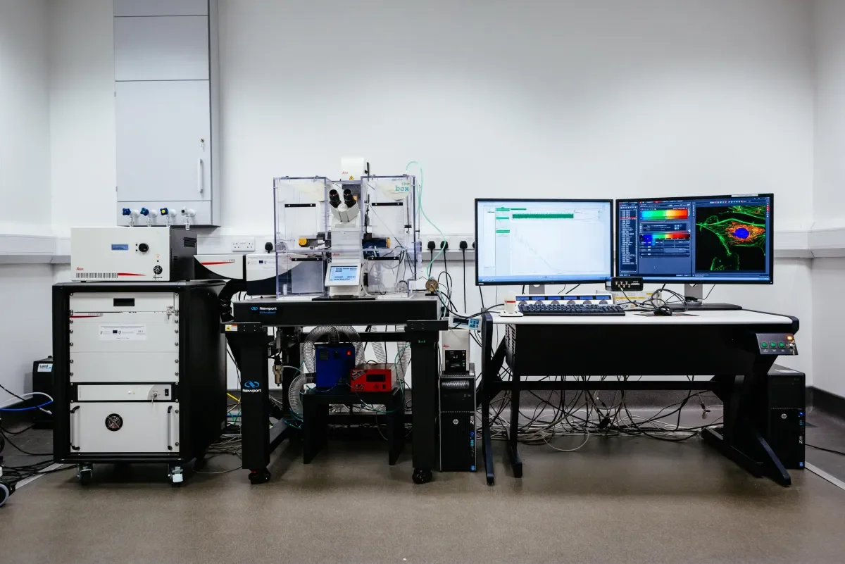
Leica TCS SP8 Confocal & STED Super Resolution Microscope
The Leica TCS SP8 STED super-resolution microscope is based on the STED (STimulated Emission Depletion) principle developed by Nobel Prize laureate Stefan Hell. The base of this system is a fluorescence confocal microscope which is suitable for use in top level biological and biomedical research and surface analysis in material science applications.
- The light sources include a pulsed 405 nm diode laser, a white light Laser which can provide excitation wavelengths from 470 nm - 670 nm, and a metal halide lamp for wide field imaging
- 592 nm and 660 nm STED depletion lasers for super resolution imaging.
- Fluorescent Lifetime Microscopy using HyD single molecule detectors
- FLIM, FCS, FLCS, FRAP and FRET measurements.
- 2 Internal standard PMT confocal detectors, 2 Internal HyD GaAsP SMD detectors for gated imaging and 1 CCD camera
- 10x air, 20x, 40x and 63x (with motorized correction collar) water, 40x, 63x and 100x oil objectives.
- Resonant scanner for imaging at 24 frames per second.
- Galvoflow, SuperZ galvanometer
- 37°C degree stage top CO² incubator for live cell imaging
- Has both high resolution and flexibility to cover a wide range of experimental setups in fluorescent microscopy
| Prices: | |
| Academic: | €29/hr |
| Industry: | On request |
| Contact: | |
| Admin: | research.facilities@dcu.ie |
| Technical: | una.prendergast@dcu.ie |
Nikon Eclipse Ti-E Fluorescence Microscope
The Nikon TiE is fully automated inverted widefield fluorescence microscope with high-speed motorization. The Eclipse Ti-E inverted microscope offers improved system speed, increased flexibility and efficient multi-mode microscopy as part of a fully-integrated microscope system that is ideal for high-end research and with its environmental control, it is perfect for live cell imaging. It has a motorised and automated XYZ, and a 37°C degree stage top CO² incubator for live cell imaging
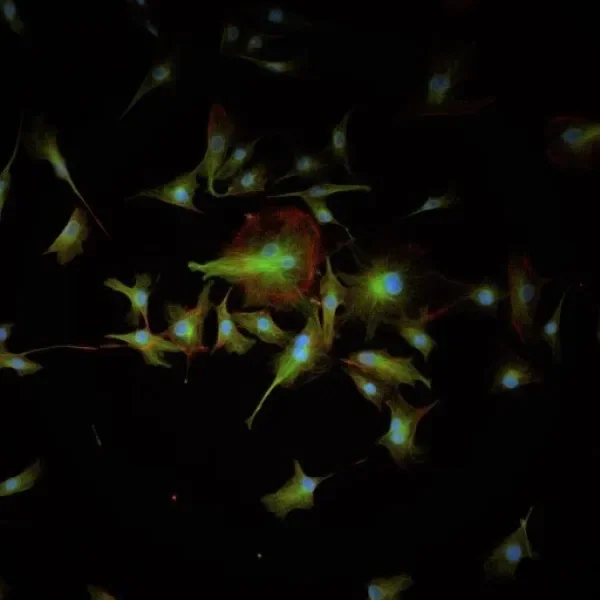
Widefield fluorescence image of mammalian cells
Imaging Modes:
-
Multichannel (λ)
-
Multipoint (x,y,λ)
-
Z Stacks (x,y,z,λ)
-
Time Series (x,y,z,λ,t)
-
Large image stitching
Objectives:
-
20x (air)
-
40x (air)
-
60x (oil)
Filter cubes:
-
DAPI
-
Alexa 488/ FITC/GFP
-
Cy3
-
Ruthenium
| Contact: | |
| Admin: | research.facilities@dcu.ie |
| Technical: | una.prendergast@dcu.ie |
Discovery Echo Revolution Widefield Microscope
The Discover Echo Revolution Microscope is a widefield fluorescence microscope that can operate in both an upright and inverted configuration, meaning a wide variety of sample types can be examined.
Sources:
- Brightfield LED Illumination
- LED Fluorescence Module
Fliter cubes:
- DAPI
- FITC
- TRITC
- TxRed
- CY5
Multi-Dimensional Imaging:
- Time-Lapse
- Multi-Point
- Mosaic
- Multi-Channel
- Z-Stack
Cameras:
- Cameras: Brightfield: 5MP CMOS Color
- Fluorescence: 5MP sCMOS Mono
Polarised and Phase contrast imaging
| Contact: | |
| Admin: | research.facilities@dcu.ie |
| Technical: | una.prendergast@dcu.ie |